Advanced microscopy offers clues to bacterial storage organelle function

Background/Objective
Bacteria lack membrane-bound organelles found in eukaryotic cells, but protein-coated compartments and other structures enable efficient coordination of cellular functions by segregating reactions or protecting fragile intermediates. Here, scientists investigated the the accumulation of carbon and inorganic phosphates in two such organelles, polyhydroxybutyrate (PHB) and polyphosphate (PP), in Rhodobacter sphaeroides.
Approach
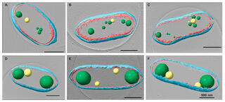
Researchers used cryo-electron tomography (cryo-ET), fluorescence microscopy, and biochemical methods to quantify size, abundance, composition, and location of PHB and PP when cell growth was arrested with chloramphenicol (Cm).
Results
Segmentation analysis and liquid chromatography and mass spectrometry revealed a 10 to 20-fold accumulation of PHB concentrated in ~1-3 granules per cell compared to ~7 granules in untreated cells. Treated cells exhibited a ~6.2-fold increase in PP granule levels, though the number of granules did not change significantly. A correlation between the location of PHB and PP granules in treated cells implies a biological advantage to the arrangement of the energy-storage organelles.
Impact
The results demonstrate the advantages of using cryo-ET to study granules and other subcellular structures. Understanding the relationship between Cm treatment and PHB or PP accumulation improves understanding of the physiological response to antibiotics. Bacterial storage compartments enriched in specific compounds could provide precursor molecules for bioplastics, biofuels, and other products.